 تويت
تويت
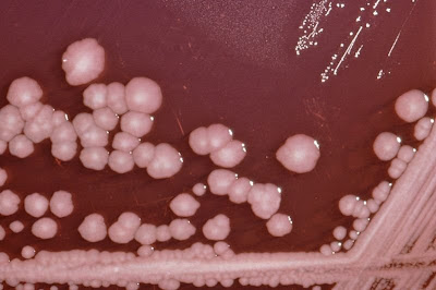
كيف اوصف pseudimonas و proteus في الآقار
وماهي الأختبارات التفريقية
وهل يوجد موقع أو كتاب يعطيني شكل شكل البكتيريا بعد تزريعها
أفيدوني جزاكم الله خيرا
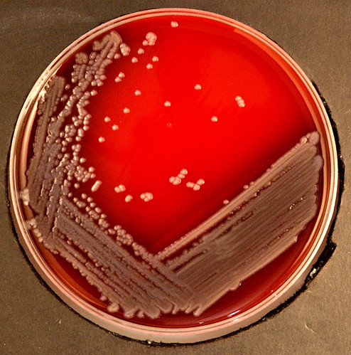
وماهي الأختبارات التفريقية
وهل يوجد موقع أو كتاب يعطيني شكل شكل البكتيريا بعد تزريعها
أفيدوني جزاكم الله خيرا


تعليق