 تويت
تويت
Streptococcus
It is Gram positive cocci in chains (groups) there is more than 100 species of streptococcus
Non motile, may be capsulated, most are facultative anaerobes but there are species that are strictly anaerobic
All are catalase negative.*
It is grown in cheep blood agar it is produced heamolysin and then it classified according to heamolysis into alfa, beta, gamma

Complete heamolysin and this strains produce*
disease ((Beta hemolytic))
disease ((Beta hemolytic))
incomplete heamolysin (partial), it formed green* around the colony ((Alfa hemolytic))
1% of streptococcus is pathogenic and 99% is commensals
The Beta haemolytic is subdivided into number of* broad groups differ in the specific polysaccharide antigens contained in the cell wall
LANCEFIELD identify a number of different group lettered in A-H and K-V
there is a special technique to identifying them is by agglutination test (as in blood group) antigen antibody binding
Streptococcus pyogen "Group A''
It is Gram positive spherical cocci, in multiplication it is plane
It is Gram positive spherical cocci, in multiplication it is plane
يعني الكريات شكلها مرتب ومصفوفة بجانب بعضها في خطوط
Unlike staphylococcus which arranged in clusters
يعني الكريات غير منتظمة وعشوائية وتشكل في مجموعات
Colony growth:
It is grown on cheep blood agar , it is appear in small colonies (pin point ) and the hemolysis is wide
It is grown on cheep blood agar , it is appear in small colonies (pin point ) and the hemolysis is wide
المستعمرة صغيرة جدا كرأس الدبوس ولكن حول هذه المستعمرة الصغيرة نجد حولها ان الدم الموجود في المزرعة قد تحلل ويظهر كدائرة كبيرة
It is grown only on cheep blood agar or chocolate agar
The S.pyogens is subdivided by specific sera into Griffith types according to their surface protein antigen (M, T and R).
Pathogenicity:
It has enzymes and toxins that enable it to invase and cause pathogenesis
streptolysins (toxins that haemolyzed red cells):
It has enzymes and toxins that enable it to invase and cause pathogenesis
streptolysins (toxins that haemolyzed red cells):
Streptolysin S
It is active aerobically ,But it is not antigenic
It is active aerobically ,But it is not antigenic
Streptolysin O
It is active under anaerobic condition ,It is antigenic stimulate the production of antistreptolysin Antibody (ASO)
It is active under anaerobic condition ,It is antigenic stimulate the production of antistreptolysin Antibody (ASO)
streptokinase: a protease that lyzes fibrin
Hyaluronidase: helps to spread in tissues by destroying hyaluronic acid, it is antigenic
leucocidin: destroys leucocytes.
lipoteichoic acid: helps in adherence to pharyngeal epithelial cells
NADase (nicotinamide adenine dinucleotidase):kills leucocytes, AB formed after infection
DNase: (deoxyribonuclease) ABCD that break
down DNA and stimulate AB response
down DNA and stimulate AB response
Erythrogenic toxin: responsible in the rash seen
in the scarlet fever and with TSST.
in the scarlet fever and with TSST.
M-proteins (antigens): anti-phagocytic virulence factors
Since it has all these factors help in phagocytosis so it cause
sore throat (tonsillitis and pharyngitis)
septicaemia
Puerperal sepsis
toxic shock syndrome (TSST)
Also hypersensitivity immune system
septicaemia
Puerperal sepsis
toxic shock syndrome (TSST)
Also hypersensitivity immune system
وهذا اهم مرض ينتج من الاصابة بهذه البكتيريا حيث ان الجهاز المناعي يبالغ في ردة فعله تجاه هذه البكتيريا وينتج عن ذلك الحاق الضرر بالعضووالأنسجة الذي تكون فيه هذه البكتيريا
Immune mediated post streptococcal infection
(AFR)Acute rheumatic fever
(AFR)Acute rheumatic fever
Will cause :-
Arthritis,Cardiaritis,Skin Rash
Arthritis,Cardiaritis,Skin Rash
(AGN) Acute Glomural Nephretitis
The test that is used to investigate streptococcus is
ASO anti-streptolysin O in serum
ASO anti-streptolysin O in serum
Laboratory diagnosis:
The specimen includes:
Throat swab
Swab of pus and serious fluid
Blood for culture
Testing ASO antibody in serum
The specimen includes:
Throat swab
Swab of pus and serious fluid
Blood for culture
Testing ASO antibody in serum
CULTURE:
BLOOD AGAR:
S.pyogens produces beta haemolytic colonies i.e. the colonies are surrounded by the zone of complete haemolysis with decolorization of the haemoglobin
Colonies are small (0.5-1 mm) colorless shiny
It is faculative anaerobe
BLOOD AGAR:
S.pyogens produces beta haemolytic colonies i.e. the colonies are surrounded by the zone of complete haemolysis with decolorization of the haemoglobin
Colonies are small (0.5-1 mm) colorless shiny
It is faculative anaerobe
Crystal violet blood agar:
It is selective medium for isolating s.pyogens
Because maybe S.aureus may be present with s.pyogenes
So crystal violet will inhibit the growth of S.Aureus
It is selective medium for isolating s.pyogens
Because maybe S.aureus may be present with s.pyogenes
So crystal violet will inhibit the growth of S.Aureus
MacConkey Agar:
S.pyogenes dose not grow on this medium.
S.pyogenes dose not grow on this medium.
BIOCHEMICALLY:
Catalase test
Catalase is an enzyme help the bacteria to protect it itself from H2O2 that produced by phagocyte
Catalase is an enzyme help the bacteria to protect it itself from H2O2 that produced by phagocyte
H2O2 ------catalaseàH2O + O
Catalase positive àStaphylococcus
Catalase negative àstreptococcus

Treatment:
S.pyogenes is very sensitive to wide range of antimicrobial drugs including penicillin and erythromycin
S.pyogenes is very sensitive to wide range of antimicrobial drugs including penicillin and erythromycin
يعالج المريض المصاب بال ِASOبالبنسلين لمدة طويلة جدا ربما لا تقل عن 15 سنة !!
Streptococcus pneumoniae
It is Gram positive diplococci, not spheric it is like two eggs held together or two flames
It forms short chains, non motile and capsulate
It is Gram positive diplococci, not spheric it is like two eggs held together or two flames
It forms short chains, non motile and capsulate
Colony morphology:
It is grown on blood agar or chocolate agar.
The colony is Alfa haemolytic
1-capsulate : it is appear mucoid
2 - non capsulated
It is grown on blood agar or chocolate agar.
The colony is Alfa haemolytic
1-capsulate : it is appear mucoid
2 - non capsulated
Pathogenisity:
S. pneumonia cause pneumonia
Meningitis
Bacteramia in HIV patients
Sever infection can occur in the elderly and in those are poorly health or immunosuppressed
Serious infection with patient of sickle cell anemia
It is normal microbial flora of the upper respiratory tract
S. pneumonia cause pneumonia
Meningitis
Bacteramia in HIV patients
Sever infection can occur in the elderly and in those are poorly health or immunosuppressed
Serious infection with patient of sickle cell anemia
It is normal microbial flora of the upper respiratory tract
Laboratory diagnosis:
The specimen can be:
Sputum
Blood for culture
CSF
The specimen can be:
Sputum
Blood for culture
CSF
Culture:
BLOOD AGAR:
Mucoid colonies, (1-2 mm) , alpha hemolytic the colonies is surrounded by partial haemolysis with green-brown discoloration in the medium.
BLOOD AGAR:
Mucoid colonies, (1-2 mm) , alpha hemolytic the colonies is surrounded by partial haemolysis with green-brown discoloration in the medium.
Chocolate agar:
It grows well on this media and the growth enhanced when incubation in co2 enriched atmosphere.
It grows well on this media and the growth enhanced when incubation in co2 enriched atmosphere.
Viridans streptococci:
These organisms are found with alpha hemolytic.
These organisms are found with alpha hemolytic.
To differentiate S.pneumoniae from other viridans there is 2 important biochemical tests:
Bile solubility
This done by emulsifying the colonies of the testing microorganism in a tube of 2 ml saline to give a turbid suspension.
This done by emulsifying the colonies of the testing microorganism in a tube of 2 ml saline to give a turbid suspension.
Then add one drop of bile (sodium deoxycholate)
Then incubate it at 37.0 C for 30 minute
If the tube becomes clear then the microorganism is S.pnumonaie
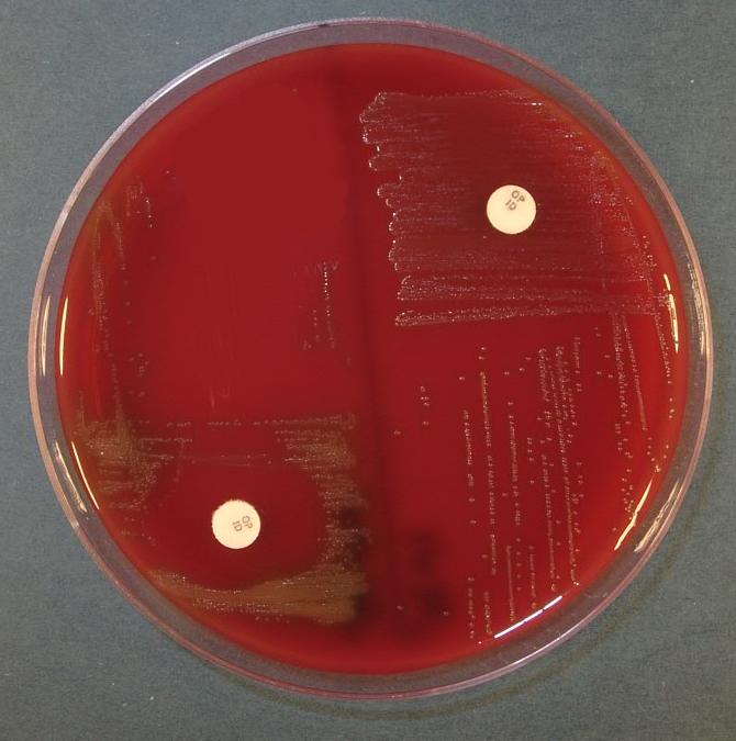
Optochin disc sensitivity
Optochin = (ethylhydrocupreine hydrochloride)
Optochin = (ethylhydrocupreine hydrochloride)
Pneumococci are sensitive to optochin
This done by a placing a disc on the culture (as antibiotic test)
Streptococcus agalactiae "Group B"
Morphology:
Gram positive streptococci in short chain, pairs and single it is discovered at 1930, non motile most are capsulated, it is B haemolysis
Morphology:
Gram positive streptococci in short chain, pairs and single it is discovered at 1930, non motile most are capsulated, it is B haemolysis
It is the major streptococcal pathogen in neonate and young infants (septcaemia and meningitis)
Pathogenicity:
It cause septic abortion, puerperal sepsis and urinary tract infection
It cause septic abortion, puerperal sepsis and urinary tract infection
It is normal flora in female genital tract
It cause neonatal septicaemia and meningitis
Laboratory diagnosis:
The specimens include:
CSF
Ear swab (it causes infection in otitis media)
Blood for culture
Vaginal swab from the women with sepsis.
The specimens include:
CSF
Ear swab (it causes infection in otitis media)
Blood for culture
Vaginal swab from the women with sepsis.
Culture:
Blood agar:
It is produced grey mucoid colonies about 2 mm in diameter,Surrounded by a small zone of B haemolysis
Blood agar:
It is produced grey mucoid colonies about 2 mm in diameter,Surrounded by a small zone of B haemolysis
MacConkey agar:
it can grow on it
it can grow on it
Kanamycin blood agar
it is a selective media
it is a selective media
Biochemical test:
1-Hippurate hydrolysis test
It is done by saline suspension of test organisms and hippurate tablet
1-Hippurate hydrolysis test
It is done by saline suspension of test organisms and hippurate tablet
2- CAMP Test
it is done by making S.aureus in the center of the blood agar and do horizontall streaking for group B without touching the S.aureus line , Arrow head will seen in the next day
it is done by making S.aureus in the center of the blood agar and do horizontall streaking for group B without touching the S.aureus line , Arrow head will seen in the next day
 >>>>>>>>>> Negative
  >>>>>>>>> Positive
It can be detected in serum or urine or CSF by using latex or coagulation slide test .this method is expensive but useful if it is not possible to isolate s.agalactiae by culturing.


 << كثرت الخيارات الغاء هذا المحرر من خيارات العضو بلوحة التحكم واستخدام المحرر العادي ..

تعليق